Drug Effects On iPSC-derived Neurons

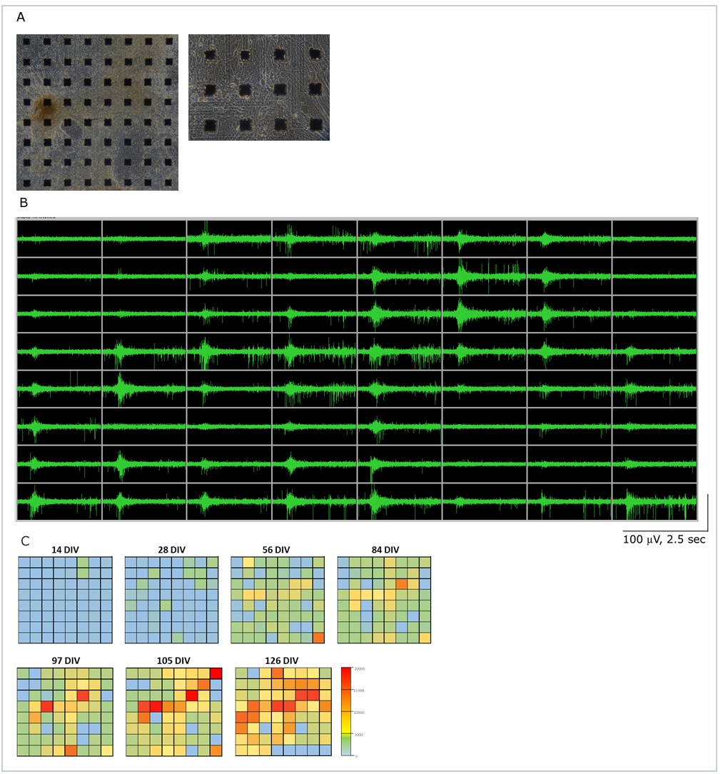
A: Human iPS cell-derived cerebral cortical neurons (Axol™ Cerebral Cortical Neurons) co-cultured with astrocyte on a MED Probe (MED-P515A, 134 days in-vitro).
B: 64 channel oscilloscope data demonstrating spontaneous activity at 97 days in-vitro.
C: Number of action potentials acquired at each of the 64 electrodes.

Movie for iPS-neurons
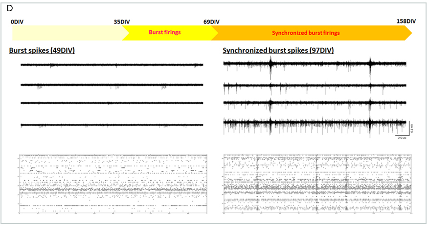
D: Comparison of waveforms and their raster plots at 49 days in-vitro and 97 days in-vitro. Burst firings were observed around at 35 days in-vitro and their synchronicity was observed around in 97 days in-vitro.

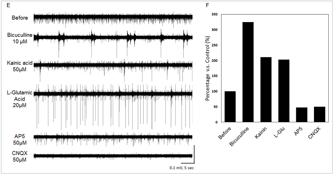
E: Spontaneous action potentials recorded from Human iPS cell-derived cerebral cortical neurons (Axol™ Cerebral Cortical Neurons) co-cultured with astrocyte . (70 days in culture). The traces represent changes in the spontaneous action potentials detected by the same electrode in response to drug application.
F: Changes of total number of spike in response to drug application.
